What Muscle Allows You to Breathe Air in and Out?
Original Editors - Rachael Lowe
Pinnacle Contributors - Khloud Shreif, Andeela Hafeez, Candace Goh, Vidya Acharya, Rachael Lowe, George Prudden, Admin, Tomer Yona, Kim Jackson, Lucinda hampton, Evan Thomas, WikiSysop, Joao Costa and Lenny Vasanthan T -
Introduction [edit | edit source]
The muscles of respiration are also chosen the 'animate pump muscles', they class a circuitous organisation in the course of semi-rigid bellows effectually the lungs.
All muscles that are attached to the man rib cage have the inherent potential to cause a breathing action.
- Muscles that helpful in expanding the thoracic crenel are called the inspiratory muscles because they help in inhalation.
- Those that compress the thoracic cavity are chosen expiratory muscles and they induce exhalation.
These muscles possess exactly the same basic structure as all other skeletal muscles, and they work in concert to expand or shrink the thoracic crenel.[1]
The speciality of these muscles are that they are composed of fatigue resistant muscle fibers, they are controlled by both voluntary and involuntary mechanisms (if nosotros want to have a breath we can, fifty-fifty if we do not think about breathing the body automatically does it)[ii]
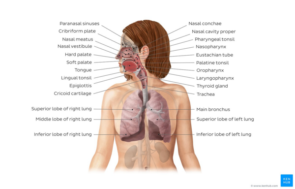
Image: Overview of the respiratory system[iii]
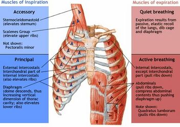
Primary Muscles [edit | edit source]
The primary inspiratory muscles are the diaphragm and external intercostals. Relaxed normal expiration is a passive procedure, happens because of the rubberband recoil of the lungs and surface tension. However there are a few muscles that assist in forceful expiration and include the internal intercostals, intercostalis intimi, subcostals and the abdominal muscles.[half-dozen]
The muscles of inspiration elevate the ribs and sternum, and the muscles of expiration depress them.[7].
Accessory Muscles [edit | edit source]
The accessory inspiratory muscles are the sternocleidomastoid, the scalenus anterior, medius, and posterior, the pectoralis major and minor, the inferior fibres of serratus anterior and latissimus dorsi, the serratus posterior superior may help in inspiration too the iliocostalis cervicis[vii]. Technically any muscle attached to the upper limb and the thoracic cage can act equally an accessory musculus of inspiration through reverse muscle activeness (musculus work from distal to proximal)[ii]
The accessory expiratory muscles are the intestinal muscles: rectus abdominis, external oblique, internal oblique, and transversus abdominis.
And in the thoracolumbar region the lowest fibres of iliocostalis and longissimus, the serratus posterior inferior and quadratus lumborum. The accessory muscles are recruited during times of exercising because of the increased metabolic need and as well during dysfunction in the respiratory organisation[6]
Diaphragm [edit | edit source]
It's a double-domed sheet of internal skeletal muscle that separates the thoracic cavity from the intestinal cavity.
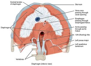
- Origin: Xiphoid process (posterior surface), lower six ribs and their costal cartilage (inner surface) and upper iii lumbar vertebrae as correct crus and upper two lumbar vertebrae every bit left crus.
- Insertion: central tendon
- Nerve Supply: Motor nerve supply by Phrenic nerve (C3 C4 C5) and sensory supply by phrenic nervus to primal tendon and lower 6 or vii intercostal nerve to peripheral parts.[8]
- Blood supply: junior phrenic arteries deliver the majority of blood supply and the remaining supply is delivered via superior phrenic, musculophrenic and pericardiacophrenic arteries.
- Action: diaphragm is the main inspiratory musculus, during inspiration it contracts and moves in an inferior direction that increases the vertical diameter of the thoracic cavity and produces lung expansion, in turn, the air is fatigued in.[9]
Intercostal muscles [edit | edit source]
They are three types: External intercostal muscles (the most superficial musculus of intercostal muscles), internal intercostal muscles, and innermost intercostal muscles.
External intercostal muscles [edit | edit source]
- Origin: inferior border of rib above and
- Insertion: superior border of rib below
Internal intercostal muscles [edit | edit source]
- Origin: from the costal groove (lower function of inner surface of rib near the inferior border) of the rib in a higher place and
- Insertion: upper border of rib beneath
Innermost intercostal muscles: [edit | edit source]
Information technology is an incomplete musculus layer and crosses more than one intercostal space. These muscles aid in the function of external and internal intercostal muscles.
- Origin: from the costal groove of the rib to a higher place and
- Insertion: the superior border of rib below
- Nerve supply: all the intercostal muscles are supplied by their respective intercostal nerves.[8]
- Claret supply: all three muscles receive claret supply from inductive and posterior intercostal arteries, in addition to internal thoracic and musculophrenic arteries; costocervical trunk for internal and innermost intercostal muscles.[ten]
References [edit | edit source]
- ↑ Breathe Strong, Perform Improve by Alison McConnell http://www.humankinetics.com/excerpts/excerpts/learn-the-anatomy-and-physiology-of-the-muscles-involved-in-breathing
- ↑ 2.0 ii.1 Pamela K. Levangie, Cynthia C. Norkin, 2005, Joint construction and function: A comprehensive analysis, 4th. Edn, Philadelphia, FA Davis publishers.
- ↑ Overview of the respiratory system image - © Kenhub https://www.kenhub.com/en/library/beefcake/the-respiratory-system
- ↑ AnatomyZone. Muscles of the Thoracic Wall - 3D Anatomy Tutorial. Available from: http://www.youtube.com/watch?five=mVLXqICrsdo[last accessed 12/4/2020]
- ↑ Armando Hasudungan. Machinery of Animate. Bachelor from: http://www.youtube.com/watch?v=GD-HPx_ZG8I[last accessed 12/4/2020]
- ↑ half dozen.0 6.1 Musles of Respiration, Wikipedia pagehttps://en.wikipedia.org/wiki/Muscles_of_respiration (accessed 30 June 2018)
- ↑ 7.0 vii.1 http://voiceandalexandertechnique.eu/voice-anatomy/pharynx-and-larynx/muscles-involved-in-vocalization-production/muscles-of-respiration.html
- ↑ 8.0 8.ane Snell's Clinical Anatomy http://teachinganatomy.blogspot.com/2013/07/respiratorymuscles.html
- ↑ TeachMeAnatomyhttps://teachmeanatomy.info/thorax/muscles/diaphragm/
- ↑ KENHUB https://www.kenhub.com/en/library/anatomy/internal-intercostal-muscles
Source: https://www.physio-pedia.com/Muscles_of_Respiration
0 Response to "What Muscle Allows You to Breathe Air in and Out?"
Post a Comment